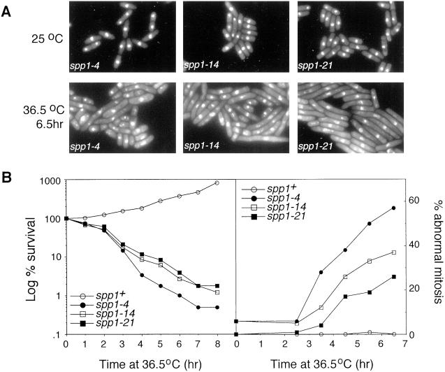
Figure 4.
Analysis of the spp1 mutants at the nonpermissive temperature. (A) Microscopic analysis of DAPI-stained spp1-4, spp1-14, and spp1-21 cells at either the permissive temperature (top) or after 6.5 h incubation at the nonpermissive temperature (bottom). (B) Survival curve of spp1 mutants at the nonpermissive temperature (left), determined by the percentage survival of cells after indicated times at 36.5°C in rich media. The percentage of cells displaying abnormal nuclear morphology (right) was determined by microscopic examination of DAPI-stained cells at the indicated times.

