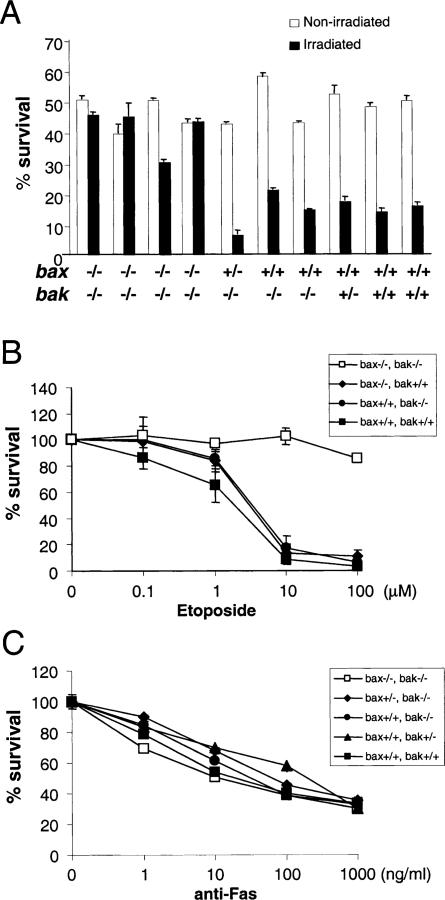
Figure 6. Thymocyte Death Assays in bax–/–bak–/– Mice.
(A) Thymocytes from bax–/–bak–/– mice and control mice were treated with 500 rad of γ irradiation (closed bars) or left untreated (open bars). Cells were stained with PI and subjected to FACS analysis after 24 hr of culture. Mean and standard deviations of triplicate samples from each mouse are presented.
(B) Thymocytes were cultured with the indicated amounts of etoposide. Cells were stained with PI and subjected to FACS analysis after 24 hr of culture. Mean and standard deviations of triplicate samples are presented.
(C) Thymocytes were cultured with indicated amounts of anti-Fas antibody. Cells were stained with PI and subjected to FACS analysis after 24 hr of culture. Mean and standard deviations of triplicate samples are presented.

