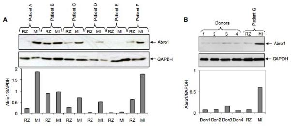
Fig. 4.

Abro1 protein levels in heart tissues from patients with coronary artery disease (CAD) and healthy donors. (A) Heart tissue extracts from six patients (A to F) with CAD were used in a Western blot to monitor the expression of Abro1 protein. Extracts were prepared from two areas on each heart that corresponded to the remote zone (RZ) and the myocardial infarction area (MI). In most patients, there was a substantial increase in Abro1 protein level in the MI area as compared to the RZ area of the same heart. The same blot was also probed with GAPDH antibody to verify equal loading. The bottom panel shows Abro1/GAPDH ratio calculated after densitometry analysis. (B) Heart tissue lysates from four healthy donors (donors 1 to 4) and one CAD patient (patient G) were used in a Western blot analysis using Abro1 antibodies. The same blot was also probed with GAPDH antibody to verify equal loading. Bottom panel shows Abro1/GAPDH ratio calculated after densitometry analysis.
