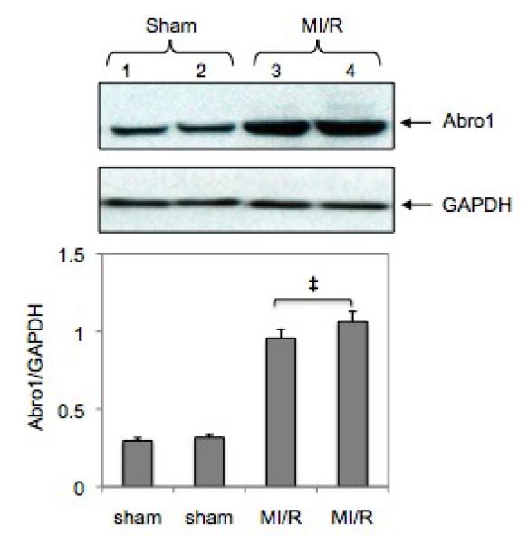
Fig. 5.

Regulation of Abro1 protein in the mouse heart following MI/R injury. Myocardial ischemia was induced for 30 minutes followed by 3 hours of reperfusion. Heart tissue extracts from sham-operated mice (n=4, two representative samples were used) or mice that underwent MI/R (n=4, two representative samples were used) were analyzed by a Western blot. There was an increase in the Abro1 protein level in the ischemic hearts (lanes 3 and 4) compared to the sham operated (lane 1 and 2). GAPDH antibody was used to verify the amount of protein present in each lane. Bottom panel shows Abro1/GAPDH ratio after densitometry analysis. Data are means ± standard deviation in animal group (n=4 hearts/group). ‡ P<0.05 vs. sham.
