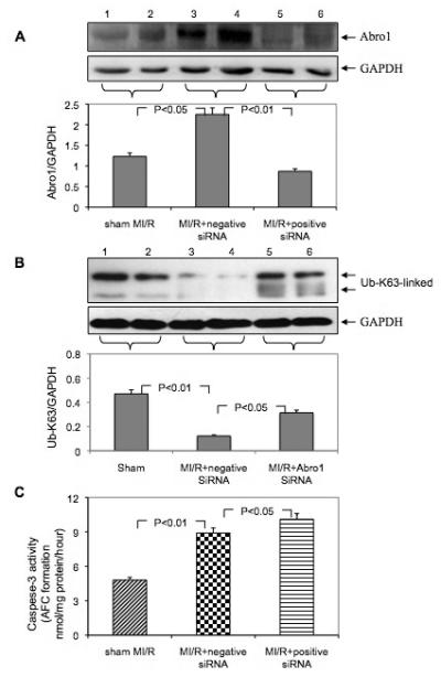
Fig. 6.

Downregulation of Abro1 protein followed by MI/R exacerbates cell injury and apoptosis. Following siRNA injection, the mice underwent MI/R, and the expression of Abro1 was monitored by Western blot analysis. (A) Western blot analysis of Abro1 protein expression from heart lysates (n=7-13 hearts/group, two representative samples of each group are presented). Lanes 1 and 2 show Abro1 protein expression in sham-operated mice. Lanes 3 and 4 show the expression of Abro1 in heart lysates from mice treated with a non-specific siRNA (negative siRNA) followed by MI/R. Lanes 5 and 6 show Abro1 expression from heart lysates in mice treated with Abro1 a specific siRNA (positive siRNA). The same blot was also probed for GAPDH expression to verify equal protein loading on each lane. The Abro1/GAPDH ratio was calculated after densitometry analysis. (B) The same heart extracts were also analyzed for K63-linked ubiquitination in MI/R+non-specific siRNA and MI/R+Abro1 specific siRNA. GAPDH antibody was used to verify the amount of protein present in each lane and the ratio of UbK63/GAPDH is shown in the bottom panel. (C) The degree of cellular damage and apoptosis was estimated by monitoring caspase-3 activity. MI/R led to a significant increase in caspase-3 activity (MI/R+non-specific siRNA) compared to control sham-operated animals. In mice that were treated with Abro1 specific siRNAs, caspase-3 activity showed a further, significant increase (MI/R+Abro1 specific siRNA). Data are means ± standard deviation in animal group (n=7-13 hearts/group).
