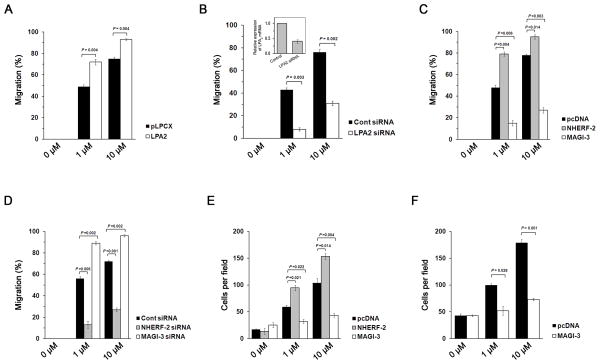
Figure 1. MAGI-3 negatively regulates cel l migration and invasion of HCT116 cells.
(A) Migration of HCT116 cells stably transfected with pLPCX or pLPCX/LPA2 in response to 1 μM or 10 μM of LPA was quantified. Full recovery of the wound was considered as 100%. (B) Migration of HCT116/LPA2 siRNA and control cells was determined. The inset shows LPA2 knockdown efficacy determined by quantitative RT-PCR. Migration of HCT116 cells (C) overexpressing NHERF-2 or MAGI-3, or (D) with knockdown of NHERF-2 or MAGI-3 by siRNA was determined. (E) Invasive capacity of HCT116/pcDNA, HCT116/NHERF-2 and HCT116/MAGI-3 cells was assessed. The cell numbers at the lower side of the invasion chamber per microscopic field were quantified. (F) Cell invasion of SW480 cells transfected with pcDNA or pcDNA/MAGI-3 was determined. n = 3 for each experimental set.

