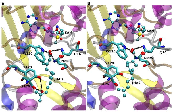
Figure 3. Docking simulations of JHA(10R) and FA.
(A) Predicted docking structure of JHA(10R). (B) Predicted docking structure of FA. Color coding is as described for figure 2. Black dotted lines represent putative hydrogen bonds between the substrates and residues Gln-14 and Trp-120, as well as between the epoxide group of JHA(10R) and Tyr-178 and Ser-176 (Panel A).

