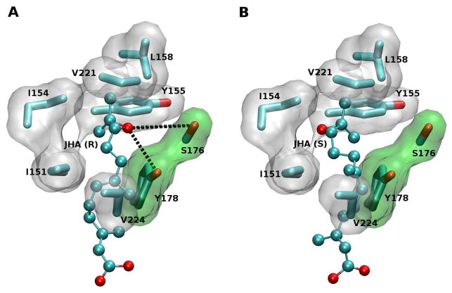
Figure 4. Epoxide recognition pocket.
(A) Close-up showing the docking of JHA(10R). The residues Ser-176 and Tyr-178 are shown with the surface colored in green, as they are both part of the pocket and have an active role in the recognition. Potential hydrogen bonds are shown in black dotted lines. The cover of the binding pocket, with the surface of the residues colored in white, is formed by the residues Ile-151, Ile-154, Leu-158, and Val-221. (B) Close-up showing the docking of JHA(10S) with the epoxide group oriented toward the hydrophobic face of the pocket (Ile-151 and Ile-154) instead of toward Ser-176 and Tyr-178.

