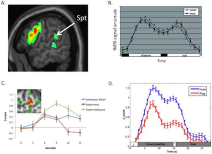
Figure 2. Location and functional properties of area Spt.
A. Activation map for covert speech articulation (rehearsal of a set of nonwords), from (Hickok and Buchsbaum, 2003). B. Activation timecourse (fMRI signal amplitude) in Spt during a sensorimotor task for speech and music. A trial is composed of 3s of auditory stimulation followed by 15s covert rehearsal/humming of the heard stimulus followed by 3s seconds of auditory stimulation followed by 15s of rest. The two humps represent the sensory responses, the valley between the humps is the motor (covert rehearsal) response, and the baseline values at the onset and offset of the trial reflect resting activity levels. Note similar response to both speech and music. Adapted from (Hickok et al., 2003)C. Activation timecourse in Spt in three conditions, continuous speech (15s, blue curve), listen+rest (3s speech, 12s rest, red curve), and listen+covert rehearse (3s speech, 12s rehearse, green curve). The pattern of activity within Spt (inset) was found to be different for listening to speech compared to rehearsing speech assessed at the end of the continuous listen versus listen+rehearse conditions despite the lack of a significant signal amplitude difference at that time point. Adapted from (Hickok et al., 2009a). D. Activation timecourse in Spt in skilled pianists performing a sensorimotor task involving listening to novel melodies and then covertly humming them (blue curve) vs. listening to novel melodies and imagine playing them on a keyboard (red curve). This indicates that Spt is relatively selective for vocal tract actions. Reprinted with permission from (Hickok, 2009b).

