Figure 1.
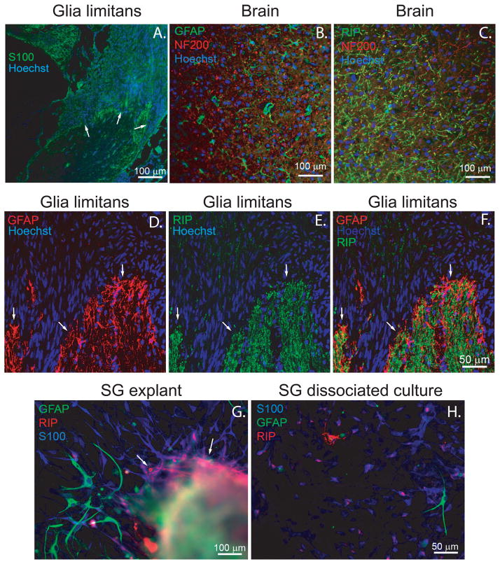
Glial cells in contact with spiral ganglion neurons and their processes express Schwann cell, astrocyte or oligodendrocyte markers. A. Frozen section of the cochlea immunostained with anti-S100 (green) and Hoechst (blue). Arrows indicate glia limitans. B&C. Frozen sections of rat cerebral cortex immunostained with anti-GFAP (green) and anti-NF200 (red) antibodies (B) or RIP (green) and anti-NF200 antibodies (C). D–F. Frozen sections of the cochlea (P60) at the glia limitans immunostained with anti-GFAP (red, D&F) and RIP (green, E&F) antibodies and Hoechst (blue). F is an overlay of GFAP and RIP immunostaining. The glia limitans is demonstrated in the intramodiolar portion of cochlear nerve and indicated by arrows. G. Area of SG explant corresponding to the intramodiolar central nerve trunk immunostained with anti-GFAP (green), anti-RIP (red), and anti-S100 (blue) antibodies. H. Dissociated SG cultures immunostained with anti-GFAP (green), RIP (red), and anti-S100 (blue) antibodies. Several GFAP-positive cells with typical astrocyte morphology migrated out from the central nerve trunk region of the SG explants (G) and composed ~2% of cells in dissociated SG cultures (H), while RIP-positive cells remained in close proximity to the central trunk region (arrows, G) and were rarely observed (<0.5% of cells) in dissociated cultures (H). Cultured SGSCs show slight RIP immunoreactivity evident as red dots on S100-positive (blue) cells (H).

