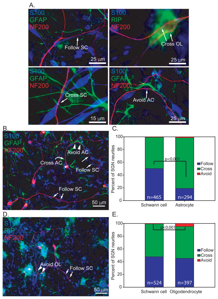
Figure 2.
Most SGN neurites extend along or cross over glial cells. Dissociated SG cultures mixed with ACs (B&C) and OLs (D&E) and immunostained with anti-NF200 (red), anti-S100 (blue), and either anti-GFAP (green) or RIP (green) antibodies. A. Neurites showed various patterns of interaction with glial cells that were classified as “follow”, “cross”, or “avoid” as illustrated here. The image showing avoidance by a nerve fiber is a higher magnification of the image in B below. B&D. Patterns of interaction with glial cells that were classified as “follow” (arrow), “cross” (curved arrow), or “avoid” (arrowhead). C. In AC-SC labeled cultures, neurites followed (50.5%) or crossed the SCs (48.8%) at nearly an equal ratio. Most neurites (79.3%) crossed ACs while 19.4% followed ACs. n=total number of processes scored. The proportion of neurites following SCs or ACs was significantly different by Chi square analysis (p<0.001) E. In OL-SC labeled cultures, 49.9% of neurites crossed OLs, 45.6% followed OLs, and a few neurites (4.5%), avoided OLs, but this was rarely observed with ACs or SCs. n=total number of processes scored. The proportion of neurites avoiding SCs or OLs was significantly different by Chi square analysis (p<0.001)

