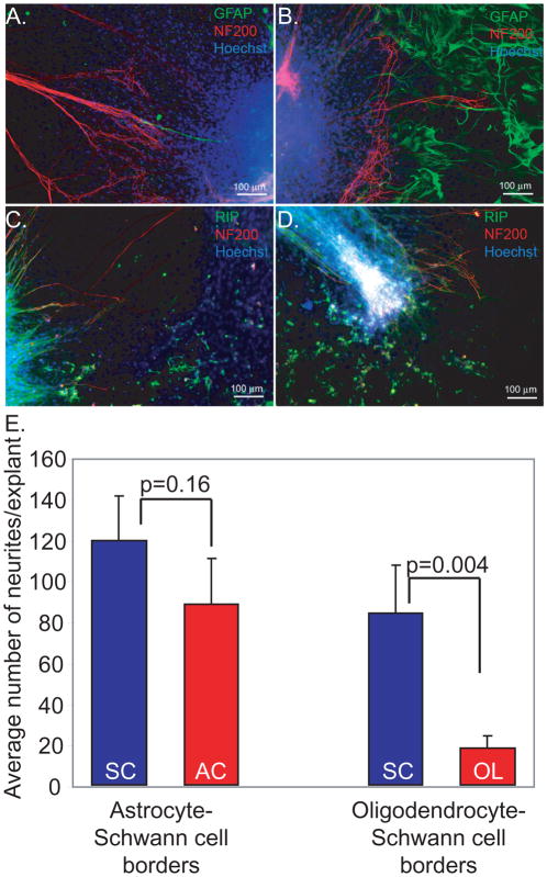
Figure 6.
More SGN neurites extend onto Schwann cells compared with astrocytes or oligodendrocytes. Cultures were immunostained with anti-NF200 (red) and either anti-GFAP or RIP (green) antibodies as indicated. Nuclei were labeled with Hoechst (blue). Regions without green colored-cells represent SCs. (A, B) Explant on the border between SCs (A) and ACs (B). (C, D) Explant on border between OLs and SCs. E. Mean number of neurites per explant plated on glial borders. More neurites grew toward SCs (119.29±21.42) than ASs (88.24±22.08) from explants placed on the border of AS-SC, but the difference was not statistically significant (p=0.16). At the border of OL-SC, significantly fewer neurites grew towards the OLs (17.70±6.18) than SCs (84.04±23.12) (p=0.004).

