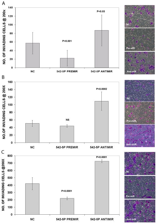
Figure 4.
Number of invading cells detected per field at 200X magnification for Kelly (A), NB1691 (B) and SKNAS (C) cells transiently transfected with PremiR-542-5p, AntimiR-542-5p and a scrambled oligonucleotide negative control (NC). Graphs represent results from two biological replicate experiments with n=3 technical repeats. All p-values are relative to the negative control. Representative images of invading cells stained with crystal violet are displayed to the right of each set of graphs.

