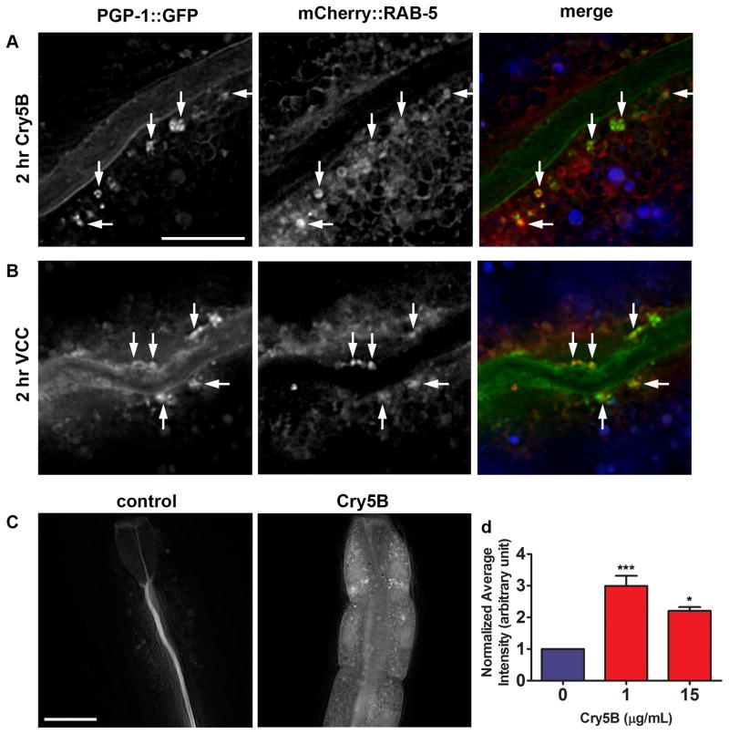
Figure 2. PFTs induce plasma membrane uptake into early endosomes and increased rates of endocytosis.
(A) PGP-1::GFP-positive vesicles induced by 2 hr exposure to E. coli-expressed Cry5B PFT and vesicles positive for mCherry::RAB-5 overlap (indicated by arrows). Due to intensity differences, overlapping signals do not always appear yellow in merged image. Autofluorescence shown in blue in the merged image. Scale bar: 10 μm. (B) Two hr exposure to V. cholerae VCC induces PGP-1::GFP-positive vesicles that show similar overlap with RAB-5::mCherry. Scale bar: 10 μm. (C) After 2 hr exposure to TRITC-labeled BSA in absence of toxin, the dye is confined to the intestinal lumen. After simultaneous exposure to 1 μg/mL purified Cry5B PFT, TRITC-BSA is abundantly found inside intestinal cells. Scale bar: 25 μm. (D) Quantification of TRITC-BSA fluorescence, in absence or presence of 1 or 15 μg/mL purified Cry5B PFT, demonstrates PFT attack leads to increased signal intensity, consistent with increased endocytosis. Means of three experiments. Error bars are standard error of the mean. Statistics indicated here and elsewhere: ns: not significant, ***: P < 0.001, *: P < 0.05.

