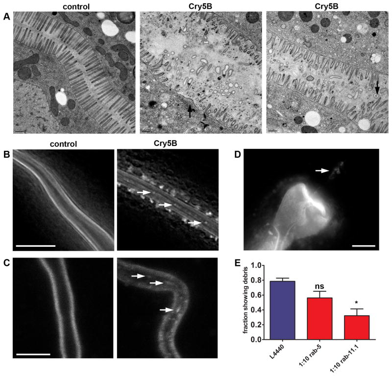
Figure 6. Cry5B PFT induces expulsion of plasma membrane into the intestinal lumen.
(A) EM images showing extensive damage to the microvilli of the intestinal cells following 3 hr E. coli-expressed Cry5B treatment. Unintoxicated controls (receptor-negative bre-5(ye17) mutant) have healthy microvilli (left). Middle and right panels: intoxicated wild type animals. Intoxicated wild-type animals show microvilli deficiency (middle, arrow), and dislodged microvilli in intestinal lumen (right, arrows). Each panel shows a single focal plane from a different animal. All focal planes were analyzed to confirm the lack of microvilli or disconnection of membranous material. Scale bars: 0.5 μM. (B) Deconvolved images showing PGP-1::GFP positive material in the intestinal lumen (arrows) after 2 hr exposure to E. coli-expressed Cry5B. Scale bar: 10 μm. (C) Confocal images showing debris in the lumen after 5 minutes exposure to E. coli-expressed Cry5B. Scale bar: 10 μm. (D) Fluorescence image showing PGP-1::GFP-labeled material in the posterior bulb of the pharynx (indicated by arrow) after exposure to E. coli-expressed Cry5B. Scale bar: 10 μm. (E) Fractions of animals containing luminal PGP-1::GFP-positive material after E. coli-expressed Cry5B PFT treatment. Error bars are standard error of the mean.

