Figure 1.
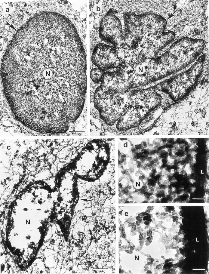
Resinless thick sections of HSF cells syringe-loaded with control bacterial extract (a and d) or HIV-1 PR at 35 μg/ml (b and e) or 70 μg/ml (c). The cells were incubated for 30 min at 37°C after syringe loading, embedded in agarose, and prepared for electron microscopy as described in MATERIALS AND METHODS. In cells treated with the control bacterial extract (a and d), the nucleus (N) is uniformly spherical or ovoid, possesses a well-defined nuclear lamina (arrow in a–c, white letter L in d and e) and is filled with an extensive and dense matrix of fibers and filaments. In cells treated with HIV-1 PR, the nuclear periphery is lobed and invaginated (b) and becomes considerably thicker than in control cells (compare L in d and e). In cells subjected to more extensive treatment with HIV-1 PR (c), the nuclear matrix is lost and the nuclear periphery becomes even more prominent, presumably due to the collapse of nuclear matrix remnants onto the lamina. Bar, 1 μm (a–c); 0.1 μm (d and e).

