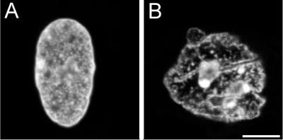
Figure 2.
Distribution of nuclear chromatin in HSF cells injected with either control bacterial extract (A) or HIV-1 PR (B). After microinjection, the cells were incubated for 15 min at 37°C and fixed; DNA was stained with propidium iodide and the preparations were subjected to CLS microscopy. Note the wholesale condensation of chromatin and multiple nuclear membrane invaginations in the HIV-1 PR-injected cell, similar to that seen in the electron micrographs of syringe-loaded fibroblasts (Figure 1, b and c). Bar, 10 μm.

