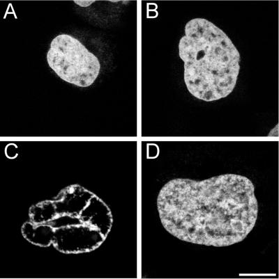
Figure 3.
Presence of the cytoplasmic IF protein vimentin correlates with the occurrence of nuclear aberrations in SW 13 cells after microinjection of the HIV-1 PR. After microinjection of either SW 13 T3 M [vimentin+] cells (A and C) or SW 13 [vimentin−] cells (B and D), the cells were incubated for 30 min at 37°C and fixed; DNA was stained with propidium iodide and the preparations were examined via CLS microscopy. (A and B) Distribution of chromatin in the cells of the cell lines SW 13 T3 M [vimentin+] and SW 13 [vimentin−], respectively, after microinjection with control bacterial extract control buffer. Equatorial optical sections of the chromatin distribution of a SW 13 T3 M [vimentin+] cell (C) and of a SW 13 [vimentin−] cell (D) after microinjection with HIV-1 PR are presented. Although the effect of the action of HIV-1 PR on nuclear chromatin organization is dramatic, the effect on nuclear shape is less obvious, partially due to the more irregular shape of the nuclei of SW 13 cells in comparison to fibroblasts. Bar, 10 μm.

