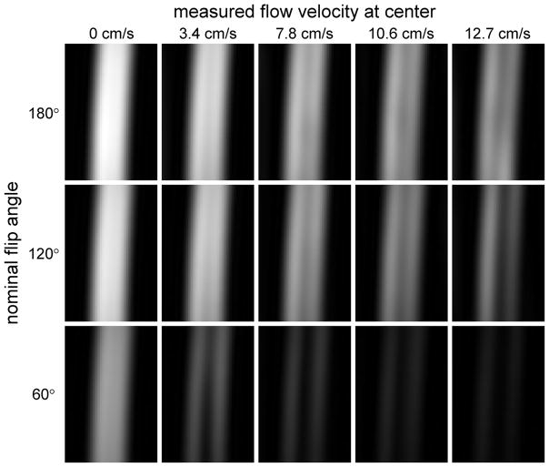
Figure 2.
Cropped images of the flow phantom acquired using the product fast spin-echo sequence with constant flip angle refocusing pulses and TE = 100ms. Images are shown for three nominal flip angles (60°, 120° and 180°) and flow rates of 0, 4, 8, 12 and 16ml/s, corresponding to measured velocities of 0, 3.4, 7.8, 10.6 and 12.7 cm/s respectively at the center of the lumen. A single slice through the center of the tube is displayed, and the same window levels are chosen for all images. Note that at zero flow the signal is lower for small flip angles. As the flow velocity increases, the signal drops faster (i.e. the flow sensitivity is greater) for small FA.

