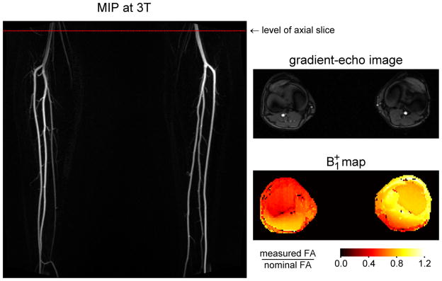
Figure 8.
Results at 3T in the same subject as Figure 1. The non-contrast MRA was acquired using an ECG-gated 3D fast spin-echo sequence with a proton density weighted variable flip angle refocusing pulse train. Note that the signal is substantially lower in the right popliteal artery than the left. Also shown is a B1+ map acquired at the level indicated by the dotted red line on the angiogram. The location of the popliteal arteries can be identified from the gradient-echo image (a magnitude image from the phase-contrast acquisition) which was obtained in the same nominal slice as the B1+ map. Note that the value of B1+ is lower at the position of the right popliteal artery than the left, supporting the hypothesis that the signal loss on the MRA is due to locally reduced effective flip angles.

