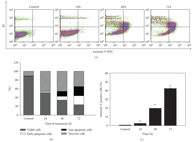
Figure 4.
Effect of LIEAF on the externalization of phosphatidylserine in Ca Ski cells. Ca Ski cells were treated without (control) or with 500 μg/mL of LIEAF for different time (24, 48, and 72 hours) and analyzed by annexin V/PI staining. (a) Flow cytometric fluorescence patterns of annexin V-PI staining. (b) Bar charts showed the percentage of distribution of viable, early apoptotic, late apoptotic and necrotic cells. (c) Results showed the percentage of annexin V positive cells. Data were means ± S.E. calculated from three individual experiments. The asterisk (*) represented significantly different from control (*P < .05).

