Figure 1.
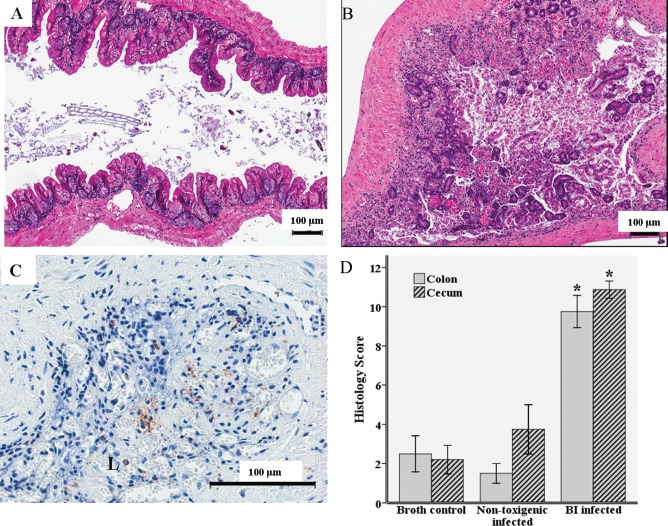
Histopathological analysis of cecal and colon segments 72 h after oral inoculation with 1 X 10⁸colony-forming units of UVA13 (BI/NAP1 [BI] strain of Clostridium difficile), VPI 11186 (nontoxigenic C. difficile strain), or broth. A, Hematoxylin-eosin stain of a cecal segment from a germ-free mouse infected with the nontoxigenic strain. B, Hematoxylin-eosin stain of a cecal segment from a germ-free mouse infected with the BI strain. C, Myeloperoxidase stain of a cecal segment from a germ-free mouse infected with the BI strain (brown indicates myeloperoxidase activity). D, Histopathological differences in the cecum and colon between BI-infected mice (N = 11), mice infected with the nontoxigenic strain (N = 2), and broth controls (N = 6) as determined by inflammatory cell infiltration, mucosal hypertrophy, vascular congestion, epithelial disruption, and submucosal edema, with a maximum score of 15. *P ⩽ .001 for BI-infected mice compared with broth controls. L, lumen.

