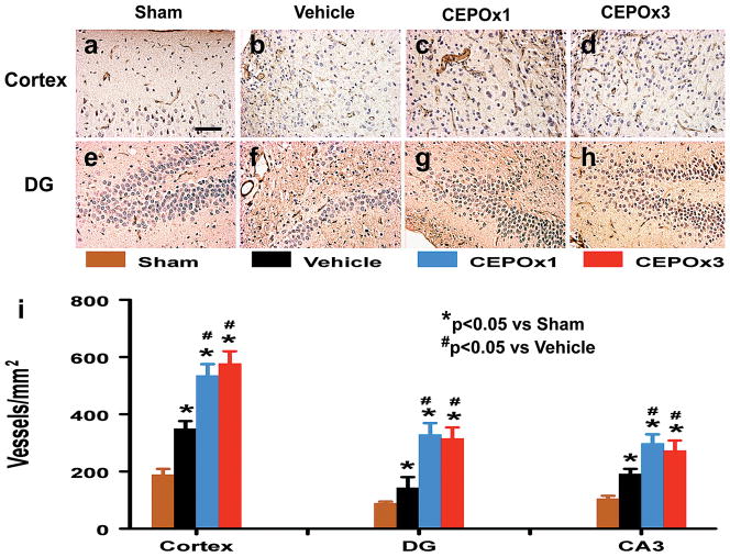
Fig. 5.
Effect of CEPO on vWF-staining vascular structure in the injured cortex and ipsilateral DG 35 days after TBI. TBI alone (b and f) significantly increases the vascular density (brown-stained) in these regions compared to sham controls (p < 0.05). CEPO treatment (c, d, g, and h) further enhances angiogenesis after TBI compared to the vehicle-treated groups (p < 0.05). The density of vWF-stained vasculature is shown in (i). Scale bar = 50 μm (a, applicable to a–h). Data represent mean ± SD. *p < 0.05 vs Sham group. #p < 0.05 vs Vehicle group. N (rats/group) = 8.

