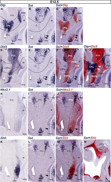
Figure 4.
Sagittal sections of E12. 5 embryos taken at lateral (A–G) and medial (H–N) levels, correlating the indicated reference markers with the presence of Sst-positive cells. The transversal boundaries of the PHy and THy are indicated by dashed lines. Note there are abundant Sst cells within basal and alar regions of the prethalamus [PTh; p3Tg in (N)], which need to be distinguished from the hypothalamic elements, found rostral to the hypothalamo-diencephalic boundary (caudal dashed line). The black arrowheads mark the incipient ventralward subpial displacement of some ABa-derived Sst cells. The white arrowheads in (G) indicate the sparse Otp-positive cells present within the CSPa subdomain. Bars = 200 μm.

