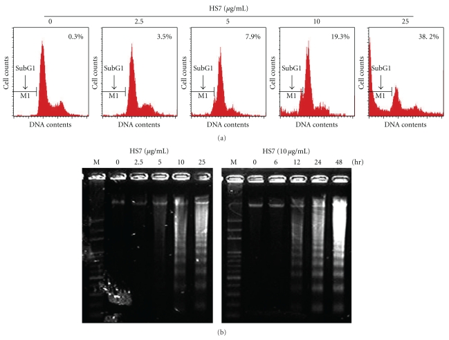
Figure 3.
Apoptosis induced by HS7 in HT29 cells. (a) HS7 increased the subG1 population in HT29 cells. HT29 cells were grown in the absence (control) or presence of HS7 (2.5–25 μg/mL) for 48 h, stained with propidium iodide (PI), and analyzed by flow cytometry for DNA content. Arrows indicate predicted location of fragmented DNA or subG1 population. (b) DNA fragmentation of HT29 cells exposed to HS7 is shown. Genomic DNA was extracted from HS7-treated HT29 cells and separated on 1.8% agarose gels.

