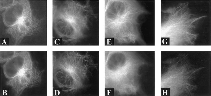
Figure 7.
Fluorescence images of living cells double-transfected with ECFP/EYFP labeled three- and/or four-repeat tau. Images were captured 2 days after transfection. (A) ECFP–tau-3R and (B) EYFP–tau-3R in cells double-transfected with ECFP–tau-3R and EYFP–tau-3R. (C) ECFP–tau-4R and (D) EYFP–tau-4R in cells cotransfected with both isoforms. Panels E–H demonstrate that interchanging the ECFP/EYFP labels does not alter the distributions of tau. (E and G) EYFP–tau-4R, ECFP–tau-4R; (F and H) ECFY–tau-3R, EYFP–tau-3R. Note: increased non–microtubule-associated tau-3R. Scale bar, 10 μm.

