
Figure 2.
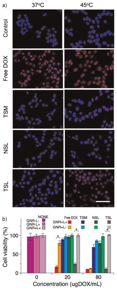
Temperature-induced intracellular drug delivery and cytotoxicity of the cooperative nanosystem. a) Images showing the intracellular delivery of DOX (red) from various therapeutic nanoparticle formulations to MDA-MB-435 human melanoma cells, at two different temperatures. The cells were incubated with free DOX, TSM, NSL, and TSL for 15 min at the indicated temperatures in a cell incubator. All formulations contain 80 μg mL−1 of DOX. Samples were rinsed three times with cell medium, then incubated for an additional 1 h at 37 °C and imaged. The nuclei were stained with 4′-6-diamidino-2-phenylindole (DAPI, blue). The scale bar indicates 100 μm. b) Cytotoxicity of various photothermally activated therapeutic nanoparticle formulations to MDA-MB-435 human melanoma cells, quantified by MTT assay (*P <0.05). The cells were incubated with free DOX, TSMs, NSLs, and TSLs at the indicated concentrations. GNR+ and GNR− indicate the presence or absence of GNRs (7 μg Au mL−1) in the mixture, respectively. L+ and L− indicate the presence or absence of NIR irradiation (810 nm, 0.75 W cm−2 for 15 min), respectively. After irradiation (or 15 min in the dark for L− samples), the cells were rinsed three times with the cell-culture medium and incubated for an additional 48 h at 37 °C before administration of the MTT assay. Values represent the mean and the error bars indicate standard deviation.