
Fig. 1.
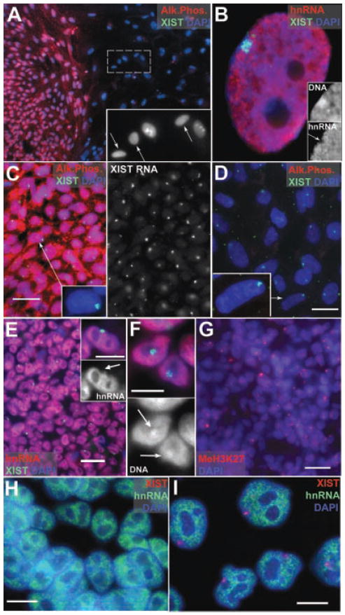
A: Undifferentiated hESC (H9) colony shows AP staining. The large differentiated nuclei among the feeders (arrows insert) do not. B: Xi in female WI38, show silencing (arrow insert), and Barr Body (insert). C: Undifferentiated hESC (ES01) with AP and XIST RA territories. D: Differentiated ES01 do not label with AP. E: Undifferentiated hESCs show a “hole” (insert, arrow) where hnRNA transcription is repressed. F: Undifferentiated hESCs have Barr Bodies (arrows DAPI channel). G: Undifferentiated colonies with MeH3K27 (red). H: Undifferentiated H9+X with no XIST RNA territories. I: Differentiated nuclei show only a single XIST territory, and hnRNA transcriptional inhibition. Scale bars: C,D,E,G,H,I = 10 μm, F and insert E = 5 μm.