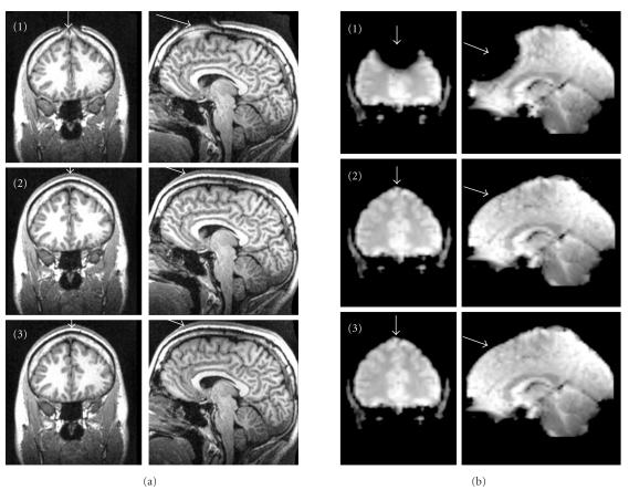
Figure 2.
(a) Structural images (MP-RAGE). (b) Functional images (EPI). Top row: standard needle (1) causing severe image distortions as well as areas of complete signal loss; middle row: non-ferromagnetic needle (2); bottom row: modified peripheral I.V. catheter (3). Both non-ferromagnetic alternatives (2, 3) did not disturb the image acquisition.

