
Figure 6. Insulin receptor regulates synapse numbers.
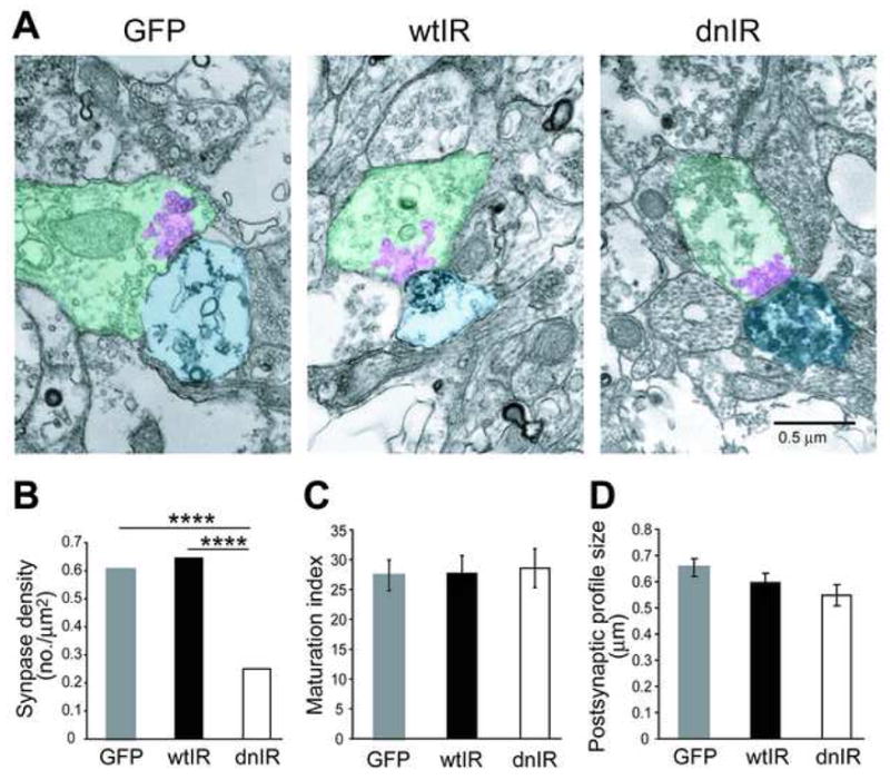
(A) Electron micrographs show ultrastructural morphology of synaptic terminals that contact GFP-, wtIR- and dnIR-expressing dendrites. Postsynaptic areas, presynaptic area and the clustered synaptic vesicle were highlighted in light blue, green and pink, respectively. (B) dnIR-expressing dendrites receive significantly fewer synapse contacts compared to GFP- and wtIR-transfected dendrites. (C) Synapses that contact GFP-, wtIR-and dnIR-expressing dendrites show comparable ultrastructural synaptic maturity, determined by the area occupied by clustered synaptic vesicles relative to the area of the presynaptic terminal. (D) GFP-, wtIR- and dnIR-expressing neurons have comparable ranges of postsynaptic profile sizes, represented by the short diameter of labeled postsynaptic area.