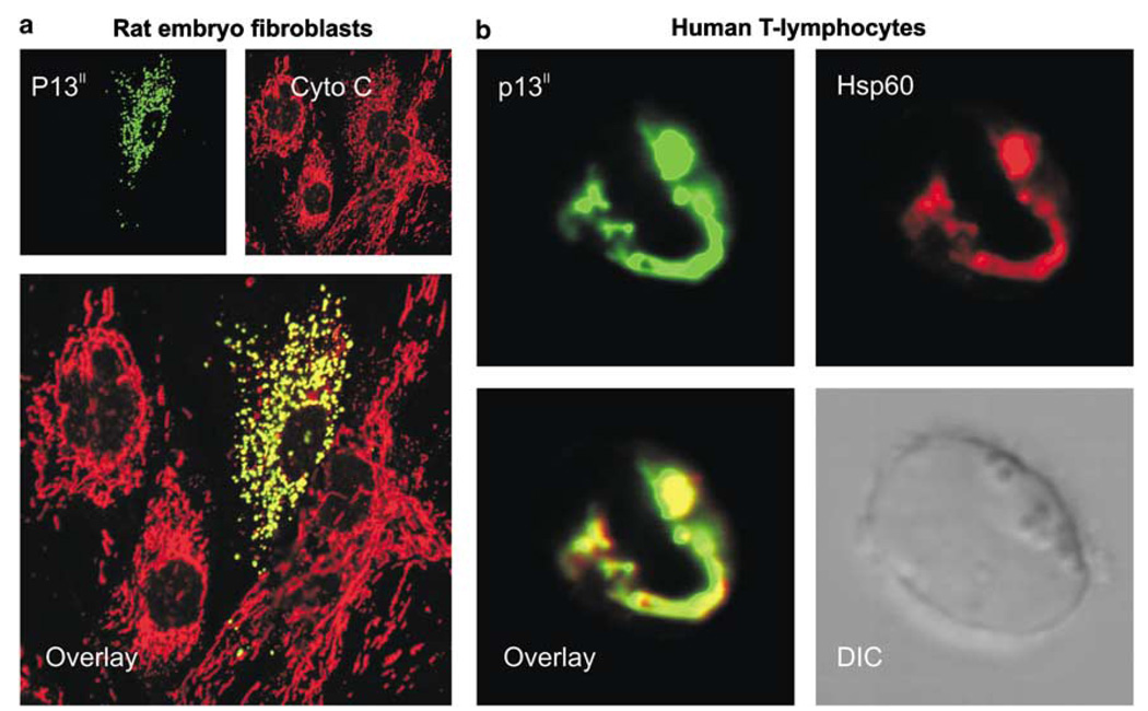
Figure 2.
Mitochondrial localization of p13II. (a) Primary rat embryo fibroblasts transfected with the p13II expression plasmid pSGp13II using Fugene 6 (Roche) are shown; (b) A human T-cell (Jurkat TetOn cell line; Clontech) transfected with the p13II expression plasmid pcDNAp13II using MegaFectin (Q.Biogene) are shown. After formaldehyde-fixation and permeabilization with Nonidet P40, the cells were analyzed by indirect immunofluorescence using a rabbit antibody recognizing p13II and Alexa 488-conjugated anti-rabbit secondary antibody (Molecular Probes); to visualize mitochondria, the cells were counterstained with mouse anticytochrome c antibody (Pharmingen; a) or goat anti-Hsp60 antibody (Santa Cruz; b) and appropriate Alexa 546 red-conjugated secondary antibodies (Molecular Probes). In addition to verifying colocalization of 13II with the mitochondrial marker proteins, (a) shows the dramatic changes in mitochondrial morphology induced by the protein

