
Figure 2. Oct4-Ctcf and Sox2-Yy1 interactions, and Tsix transcriptional activation.
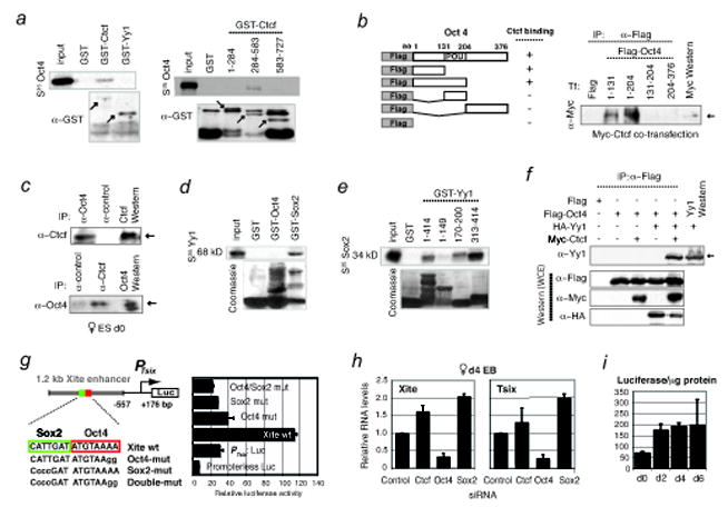
a, Testing mammalian GST fusions of full-length Yy1 and Ctcf or Ctcf domains (amino acids indicated) for interaction with S35-labelled Oct4. Lower panels, α–GST Western control. Arrows, specific protein fusions.
b, HEK cells were cotransfected with myc-Ctcf and indicated Flag-tagged Oct4 fragments. Whole cell extracts (WCE) were immunoprecipiated (IP) with α-Flag antibodies prior to Western blotting with α-myc antibodies. Arrow, Ctcf protein.
c, Reciprocal co-IP: Left, IP with α-Oct4 or control antibodies to test interaction with endogenous Ctcf; arrow, Ctcf detected by α-Ctcf Western analysis. Right, IP with α-Ctcf or control antibodies to test interaction with endogenous Oct4; arrow, Oct4 detected by α-Oct4 Western. Doublet bands and mobility differences may reflect isoforms due to post-translational modifications.
d, Yy1 (S35-labelled) binding to Sox2 but not Oct4 revealed by GST fusion analysis. Bottom, Coomassie staining to reveal GST fusion proteins.
e, Domain mapping of Yy1 binding to Sox2. Mobility differences may reflect post-translational modifications.
f, Overexpression and co-IP of Flag-Oct4, Myc-Ctcf, and HA-Yy1 in indicated combinations in HEK cells. Two 6-well plates of HEK cells were transfected with 8 μg of tagged constructs and harvested 35 hrs later for coIP analysis. Western blots of the co-IP (top panel) and whole cell extracts (bottom 3 panels) were performed using indicated antibodies.
g, Oct4 and Sox2 confer developmental specificity to Xite enhancer. Map of luciferase expression vector fused to the Tsix major promoter (PTsix) and the 1.2 kb enhancer. Mutations of the Oct4/Sox2 motifs in the enhancer compromised PTsix activity in transiently transfected male d0 ES cells. Error bars, 1 SD.
h, qRT-PCR for Xite and Tsix RNA after knocking down the indicated factors. Levels are normalized to β-actin levels. Error bars, 1SD.
i, Luciferase analysis of cells carrying the wildtype enhancer shows enhanced PTsix activity upon differentiation. Cells were differentiated in duplicate and luciferase levels were normalized to total protein levels.