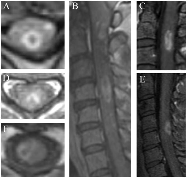
Figure 1. Spinal cord ring enhancement patterns in multiple sclerosis.

A. Case 1: Axial T1-weighted imaging post gadolinium (TR=908, TE=14) demonstrates closed ring enhancement at the level of C4. B. Case 1: Sagittal T1-weighted imaging post gadolinium (TR=828, TE=9.7) reveals multiple ring enhancements a different levels (C1 and C4) and within the same lesion (C4). C. Case 2: Sagittal T1-weighted imaging post gadolinium (TR=500, TE=14) shows a ring extending longitudinally and opening superiorly at the level C2-3. D. Case 3: Axial T1-weighted imaging post gadolinium (TR=615, TE=11) demonstrates ring enhancement opening out dorsally at the level C4. E. Case 14. Sagittal T1-weighted imaging post gadolinium (TR=600, TE=16.4) shows open ring enhancement opening out dorsally at the level C4-5. F. Case 17. Axial T1-weighted imaging post gadolinium (TR=460, TE=14) demonstrates ring enhancement opening toward the center of the cord at the level C2.
