FIGURE 1:
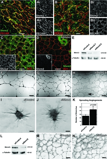
Shrm2 knockdown stimulates endothelial angiogenesis. (A and B) Murine yolk sacs at E9.5 stained with PECAM and Shrm2 antibodies show Shrm2 localization at cellular junctions in both large vessels (A) and the capillary plexus (B). (C and D) C166 endothelial cells were treated with a nontargeting siRNA (siControl) (C) or a Shrm2-specific siRNA (siShrm2) (D) and were stained with Shrm2 (inset) and ZO1. (E) Shrm2 knockdown from two different siRNAs was confirmed via Western blot. α-Tubulin was used as a loading control. (F–H) siControl (F), siShrm2–7 (G), or siShrm2–8 (H)–treated C166 cells were plated on matrigel to examine angiogenic potential. Arrow indicates an area that has failed to undergo angiogenesis. (I–K) siControl (I) and siShrm2 (J) C166 cells were grown as spheroids for use in a collagen sprouting angiogenesis assay. Quantification of collagen sprouting angiogenesis is shown in (K). The numbers of branch tips are represented as the mean ± SD (n = 7 spheroids). (L) Western blot of Shrm2 knockdown in HUVECs. (M and N) Matrigel angiogenesis assay for siControl (M) and siShrm2 (N)–treated HUVECs. Scale bars = 25 μm in A–D; 1 mm in F–H; 125 μm in I, J, M, and N.

