FIGURE 5:
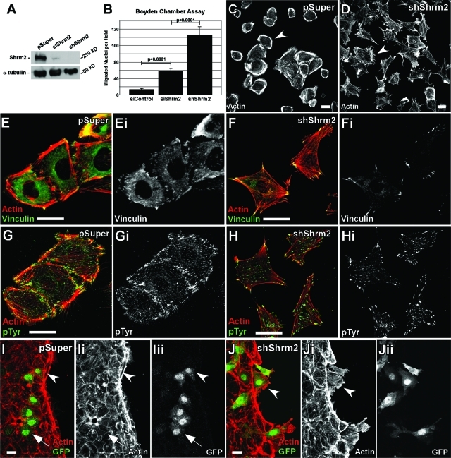
Stable expression of shShrm2 changes endothelial morphology and enhances migration. (A) Western blot of transient siShrm2, stable shShrm2, or stable vector control (pSuper) C166 cells for Shrm2. α-Tubulin was used as a loading control. (B) Quantification of Boyden chamber migration for siControl, siShrm2, or shShrm2 C166 cells. The number of migrated nuclei is represented by the mean ± SEM (n = 6). (C and D) pSuper (C) or shShrm2 (D) C166 cells were allowed to spread on fibronectin-coated coverslips for 4 h and were immunostained for actin. Arrowheads indicate differences in cortical actin. (E–H) pSuper (E and G) or shShrm2 (F and H) C166 cells were immunostained for vinculin (E and F) or phospho-tyrosine (G and H) to visualize focal adhesions. (I and J) pSuper (I) or shShrm2 (J) C166 cells (indicated by GFP) were mixed with parent C166 cells and wounded. Cells were allowed to migrate for 30 min and were then stained for GFP and actin. Arrows indicate stable junctions formed between pSuper and parent C166 cells. Arrowheads indicate pSuper cells that contribute to the actin belt or shShrm2 cells that migrate quickly into the wound past the actin belt. Scale bars = 25 μm.

