FIGURE 6:
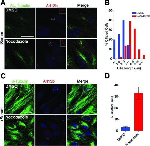
Nocodazole-mediated microtubule depolymerization affects cilia length. (A) Forty-eight-hour serum-starved htRPE cells were treated with 100 nM nocodazole for 2 h and stained with antibodies to anti-acetylated α-tubulin (green) and anti-Arl13b (red). Scale bar 15 μm and nuclei stained blue with Hoechst. (B) Quantification of cilia length distribution in nocodazole-treated cells (from A). Mean cilia length for 100 nM nocodazole (3.9 ± 0.14) and DMSO (1.9 ± 0.15). Values as mean ± SEM. (C) htRPE cells in high-serum growth media were treated with 100 nM nocodazole for 2 h. Cells were stained with anti–β-tubulin (green) and anti-Arl13b (red) antibody to visualize microtubules and cilia. Scale bar 15 μm and nuclei stained blue with Hoechst. (D) Quantification of percentage of ciliated cells (from C). P < 0.001.

