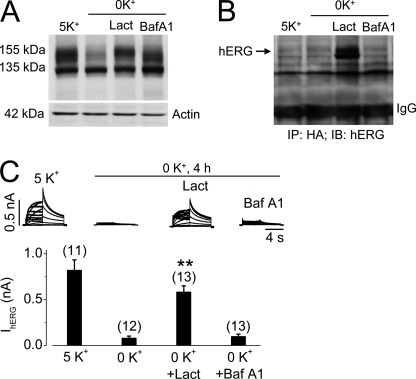
FIGURE 6.
Effects of proteasomal or lysosomal inhibition on 0 mm K+-induced hERG internalization in UbKO (HA-tagged)-transfected hERG-HEK cells. A, Western blots showing hERG expression levels in UbKO-transfected hERG-HEK cells cultured for 4 h in 5 mm K+ MEM, 0 mm K+ MEM, 0 mm K+ MEM with 20 μm lactacystin (Lact), or 1 μm bafilomycin A1 (Baf A1). B, co-IP analysis between expressed UbKO and hERG channels. Proteins extracted from UbKO-transfected hERG-HEK cells cultured for 4 h in 5 mm K+ MEM, 0 mm K+ MEM, 0 mm K+ MEM with 20 μm lactacystin (Lact), or 1 μm bafilomycin A1 (Baf A1) were precipitated using an anti-HA antibody. The precipitated proteins were immunoblotted (IB) using an anti-hERG antibody. C, effects of lactacystin or bafilomycin A1 on 0 mm K+-induced reduction of IhERG. Families of IhERG were recorded in hERG-HEK cells cultured in 5 mm K+ MEM, 0 mm K+ MEM, 0 mm K+ MEM with 20 μm lactacystin (Lact), or 1 μm bafilomycin A1 (Baf A1) for 4 h. Cells were collected in drug-free 5 mm K+ MEM, and IhERG was recorded using the whole cell clamp method in 5 mm K+ bath solution. Tail current at −50 mV after a 50-mV depolarization was used for analysis. Currents from cells cultured in 0 mm K+ MEM with lactacystin (Lact) or bafilomycin A1 (Baf A1) were compared with currents from cells in 0 mm K+ MEM. The numbers in parentheses denote the number of cells tested. Error bars represent standard error of the mean. **, p < 0.01 compared with IhERG in 0 mm K+.

