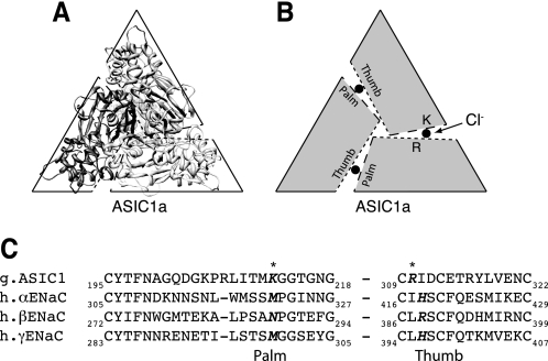
FIGURE 1.
Sequence alignment of ASIC1a with α-, β-, and γENaC. A, ribbon structure of chicken ASIC1a (2QTS) shown perpendicular to the plane of the membrane from an extracellular perspective. B, model of ASIC1a subunit arrangement. Dotted lines represent the thumb domain, dashed lines represent the palm domain. Approximate location of Cl− is depicted by a filled circle located between the thumb and palm domains of two adjacent subunits. C, partial sequence alignment of the palm and thumb domains of chicken ASIC1a with human α-, β-, and γENaC using the ClustalW method. ASIC1a Cl− coordinating residues are indicated with an asterisk. Potential ENaC Cl−-coordinating residues identified by sequence alignment are in bold.

