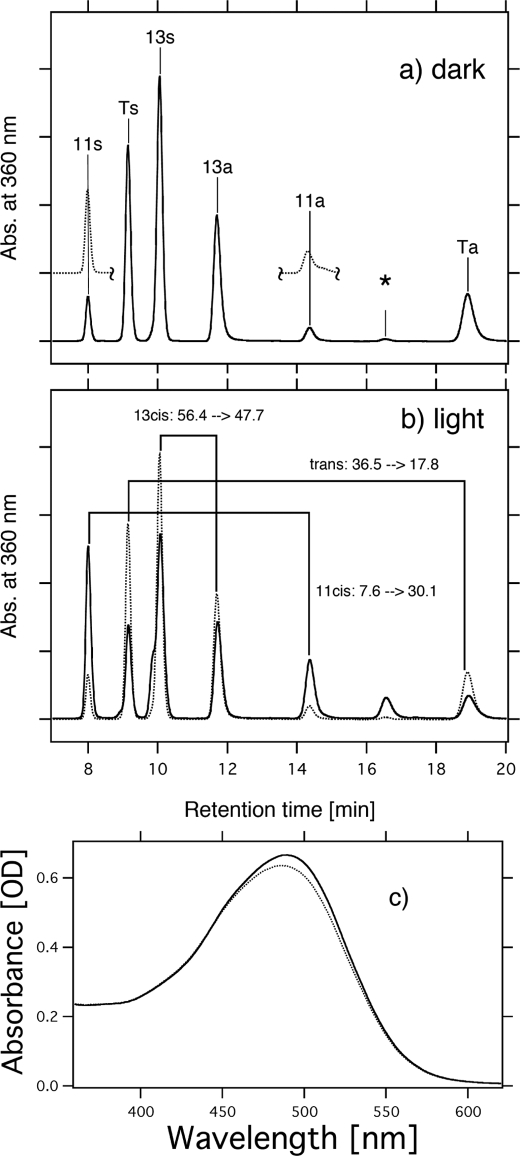
FIGURE 3.
Conformation of retinal extracted from MR in 1 m NaCl, 0.05% DDM, and 50 mm Tris-HCl, pH 7.0, in the dark (a) and upon illumination with >460 nm light for 10 min (b). The detection beam was set at 360 nm. Ts, Ta, 11s, 11a, 13s, and 13a stand for all-trans-15-syn-retinal oxime, all-trans-15-anti-retinal oxime, 11-cis-15-syn-retinal oxime, 11-cis-15-anti-retinal oxime, 13-cis-15-syn-retinal oxime, and 13-cis-15-anti-retinal oxime, respectively. The HPLC pattern of 11-cis-retinal obtained by irradiating all-trans-retinal is shown as dotted lines (a). One division of the y axis of a and b corresponds to 5000 absorbance units. An asterisk means an unknown peak, presumably originating from 9-cis-retinal oxime. The molar composition of retinal isomers was calculated from the areas of the peaks in the HPLC patterns. c, absorption spectra measured in the dark (dotted line) and upon illumination with >460 nm light for 10 min (solid line).

