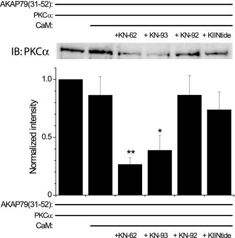
FIGURE 4.
Classical CaMKII inhibitors induce apoCaM-mediated displacement of PKC from AKAP79. AKAP(31–52) was incubated with PKCα (200 ng) in the absence or presence of apoCaM (10 μm) and various CaMKII reagents (each 1 μm). Top, representative Western blot of PKCα binding to AKAP79(31–52) in the absence or presence of apoCaM and the indicated CaMKII reagents. Bottom, graph summarizing the data (mean ± S.E.) from multiple experiments (n = 6). For each experiment, the data were normalized to the amount of PKC bound to AKAP79(31–52) alone. *, p < 0.05; **, p < 0.01 compared with PKCα + AKAP79(31–52) + apoCaM. KIINtide, CaMKIINtide; IB, immunoblot.

