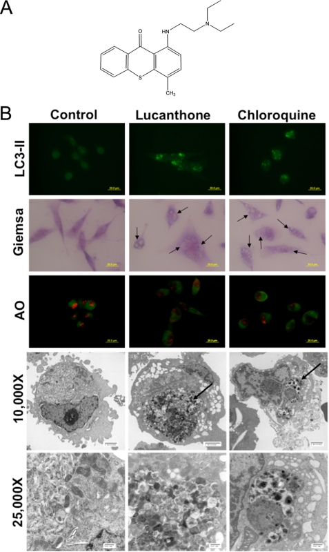
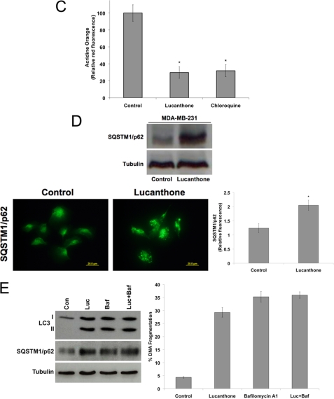
FIGURE 1.
Lucanthone inhibits autophagy. A, chemical structure of lucanthone. B, lucanthone induces LC3-II formation, vacuolization, and LMP. MDA-MB-231 cells were treated for 48 h with 10 μm lucanthone or 50 μm chloroquine. LC3-II was visualized by immunocytochemistry, vacuolization by Giemsa staining, and LMP by loss of acridine orange fluorescence. Electron microscopy demonstrates vacuolization and electron dense particle accumulation. Arrows indicate undegraded protein accumulation. C, quantification of LMP. The degree of red acridine orange staining was measured in MDA-MB-231 cells by immunofluorescence and quantified using ImageJ software. D, lucanthone stimulates SQSTM1/p62 accumulation and aggregation. Cells were treated with 10 μm lucanthone for 48 h. Levels and aggregation of p62 were determined by immunoblotting and immunocytochemistry, respectively. Fluorescence was quantified using ImageJ software. Mean ± S.D., n = 5. * indicates a significant difference from the controls. p < 0.05. E, Bafilomycin A1 does not augment lucanthone-mediated p62 or LC3-II accumulation or apoptosis. MDA-MB-231 cells were treated with 10 μm lucanthone, 100 nm bafilomycin A1, or both agents for 48 h.


