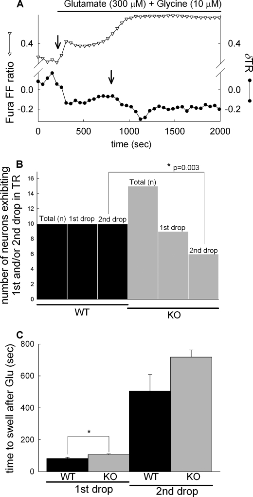
FIGURE 6.
Effect of cypD ablation on glutamate-evoked biphasic mitochondrial swelling in cortical neurons. A, wide field fluorescence time lapse recording in a fura-FF/AM-loaded WT cortical neuron expressing mito-DsRed. Glutamate (300 μm) plus glycine (10 μm) in the absence of [Mg2+]e evoked a rise of [Ca2+]i indicated by the increasing ratio of fura-FF 340/380-nm fluorescence intensities (triangles). Mitochondrial swelling was measured by calculating TR of mito-DsRed fluorescence images (circles), where swelling is marked by the decreasing TR (arrows). The first and second drops of the TR always coincided with the initial response to glutamate and to the delayed Ca2+ deregulation, respectively. Representative traces of 10 recordings are shown. B, quantification of the observation of first and second drops in neurons from WT (black bars) and cypD-KO mice (gray bars) using Fisher's exact test. Total (n) corresponds to the number of cells observed and first and second drops to the observation of the drops in the TR in recordings similar to A. C, onset of mitochondrial swelling defined as the time elapsed between the application of glutamate and the sudden decrease of the TR; bars show mean ± S.E. (error bars) of 10 cells (*, p < 0.05 significance by Kruskal-Wallis ANOVA on Ranks).

