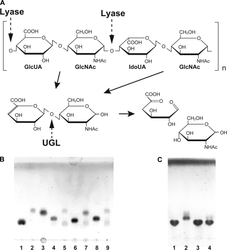
FIGURE 1.
UGL reaction. A, scheme of heparin degradation by UGL. The heparin molecule contains GlcUA and IdoUA. Dotted and solid arrows indicate the cleavage sites for polysaccharide lyases and UGL and the degradation pathway for polysaccharides, respectively. B, unsaturated glycosaminoglycan disaccharides were incubated at 30 °C with SagUGL, and the resultant products were detected on the TLC plate. Lane 1, GlcUA (70 nmol); lane 2, GlcNAc (70 nmol); lane 3, GalNAc (70 nmol); lane 4, unsaturated heparin disaccharide (52.5 nmol); lane 5, unsaturated heparin disaccharide (52.5 nmol) with SagUGL; lane 6, Δ0S (52.5 nmol); lane 7, Δ0S (52.5 nmol) with SagUGL; lane 8, unsaturated hyaluronan disaccharide (52.5 nmol); and lane 9, unsaturated hyaluronan disaccharide (52.5 nmol) with SagUGL. C, Δ4S (150 nmol) was incubated at 30 °C with SagUGL or BacillusUGL, and the resultant products were detected on the TLC plate. Lane 1, Δ4S (150 nmol); lane 2, Δ4S (150 nmol) with SagUGL; lane 3, Δ4S (150 nmol) with BacillusUGL WT; and lane 4, Δ4S (150 nmol) with BacillusUGL H210R.

