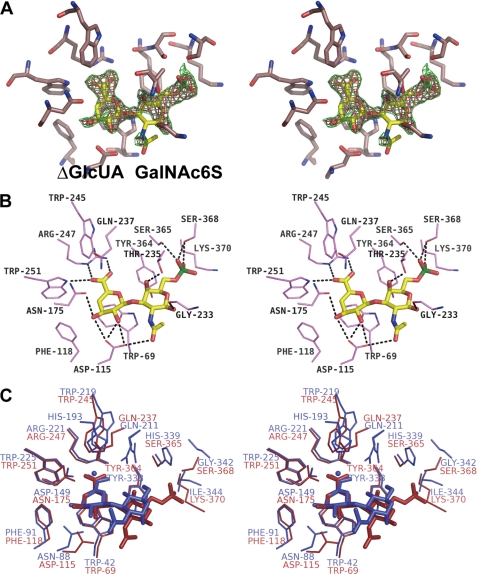
FIGURE 3.
Active site structure of SagUGL D175N in complex with Δ6S. A, electron density of Δ6S in the omit (Fo − Fc) map calculated without the substrate and contoured at the 2.5 (green) and 3.0 σ (red) levels. B, interaction of SagUGL D175N with Δ6S (stereodiagram). Several residues bind to Δ6S through the formation of hydrogen bonds (broken lines). Atoms carbon, oxygen, nitrogen, and sulfur of Δ6S are colored yellow, pink, blue, and green, respectively. C, superimposition of SagUGL D175N-Δ6S complex (red) and BacillusUGL D88N-Δ0S complex (blue).

