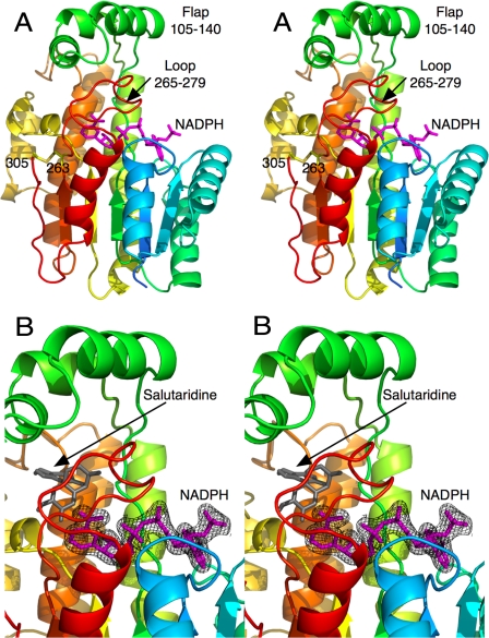
FIGURE 1.
Structure of SalR. A, stereo image of the structure of SalR looking into the active site. The ribbon is colored from blue to red as the amino acid chain extends from the N to the C terminus. The structure of the bound NADPH is represented by a mauve stick figure. B, close-up of the active site showing the electron density of the bound NADPH molecule. As a reference, the modeled salutaridine is shown as a gray stick figure.

