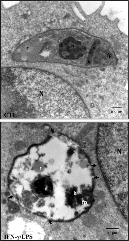
FIGURE 5.
Electron microscopy studies of T. cruzi infection. Micrographs showing unstimulated (CTL) and activated (IFN-γ/LPS) infected macrophages at 1 h post-infection. The arrows in the lower panel indicate disruptions of membrane integrity. N, macrophage nucleus; T. cruzi n, T. cruzi nucleus; k, kinetoplast; fp, flagellar pocket; r, reservosomes. The electron micrographs are representative of at least three independent experiments.

