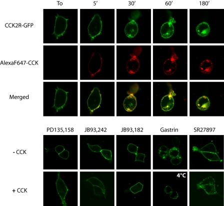
FIGURE 2.
Confocal microscopy imaging of stimulated CCK2R internalization in HEK cells. HEK cells were transfected with CCK2R-GFP and were incubated at 37 °C with Alexa F 647-CCK for increasing times (upper panels) or with different pharmacological agents for 30 min: partial agonists of the CCK2R on inositol phosphate turnover (PD135,158 and JB93,242, 10 μm), an inverse agonist (JB93,182, 10 μm), the full agonist of the CCK2R (gastrin, 0.1 μm), or an antagonist of the CCK1R (SR27,897, 10 μm) alone or in combination with 0.1 μm CCK (lower panels). The images show internalization of CCK2R-GFP following stimulation by full agonists CCK and gastrin but not other compounds, including potent partial agonists on inositol phosphate turnover that behave as antagonists on CCK2R internalization. Merged images show co-localization of Alexa F 647-CCK with CCK2R-GFP over time. Images are representative of at least three separate experiments.

