
Figure 2. Snapshots from simulations of various hybrid models of tumor growth.
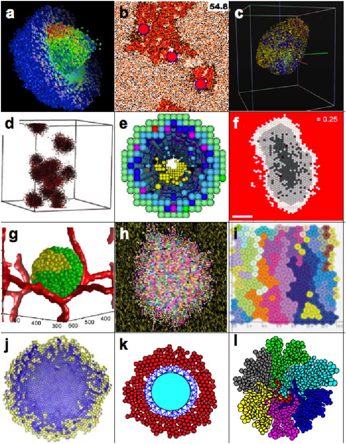
a) 3D tumor spheroid, simulated by a hybrid cellular automaton; reprinted from [8] with permission from Birkhauser-Verlag. b) Tumor invasion in prostate ducts simulated by a hybrid cellular automaton; Reprint permission requested from [16]. c) 3D tumor spheroid simulated by an agent-based on-lattice model; reprinted from [10] with permission from Springer. d) 3D tumor self-metastatic spheroids simulated by a hybrid cellular automaton; reprinted from [5] with permission from Nature Publishing Group. e) 3D model of ductal carcinoma in situ simulated by a square-grid cellular automaton; reprinted from [18] with permission from Elsevier. f) 2D tumor spheroids simulated by a hexagonal cellular automaton; reprinted from [34] with permission from BioMed Central, the Open Access Publisher. g) 3D vascularized tumor spheroid simulated by Potts model; reprinted from [46] with permission from Public Library of Science, open access article. h) 2D tumor spheroid in a heterogeneous environment composed on ECM fibers simulated by Potts model; reprinted from [42] with permission from Elsevier. i) 2D model of colorectal tumor simulated using the particle model with Voronoi triangulation; reprinted from [61] with permission from John Wiley and Sons. j) 2D tumor spheroid modeled using the cell-centered off-lattice model; reprinted from [50] with permission from Springer. k) 2D hybrid model of tumor growth simulated by particle center-based ellipsoid model; reprinted from [56] with permission from World Scientific. l) 2D multiclonal tumor growth simulated by a model of deformable fluid-based cells; reprinted from [69] with permission from Birkhauser-Verlag.