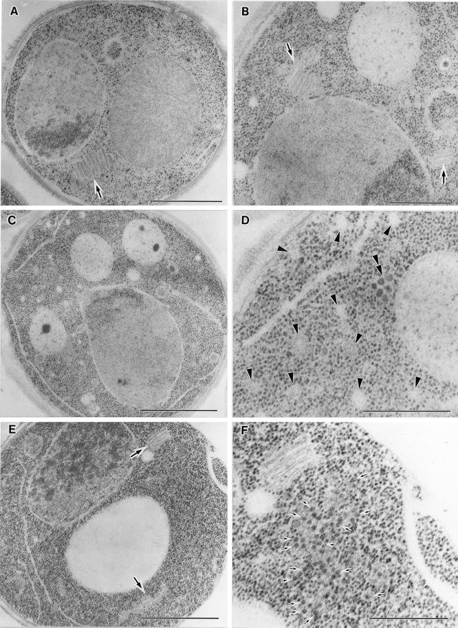
Figure 10.
Electron microscopic observation of the arf1-18 ts mutant. arf1-18 ts mutant cells were grown to an early log phase at 23°C, further incubated at 23°C (A) or at 37°C for 1 h (B, C, and D) and 2 h (E and F), and then subjected to electron microscopy. In A, B, and E, large arrows mark Golgi structures. In D and F, various kind of vesicles are indicated by arrowheads, a double arrowhead, and small arrows. Bars: A, C, and E, 1.0 μm; B, D, and F, 500 nm.

