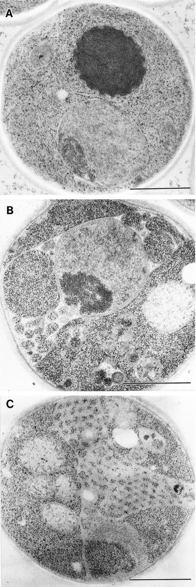
Figure 6.

Electron microscopic observation of arf1-11 ts mutant cells. Wild-type (YPH499, WT) (A) and arf1-11 ts mutant cells (B and C) were grown to an early log phase at 23°C, incubated at 37°C for 1 h (B) and 4 h (A and C), prepared for electron microscopy by the freeze-substitution fixation method, and observed under an electron microscope. Bars, 1.0 μm.
